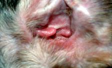
HEMATOMA OF THE EAR
An aural (ear) hematoma is a collection of blood, serum, or a blood clot within the pinna (ear flap).
When present, the pinna will appear very thick and spongy. The swelling may involve the entire
pinna or it may involve only one area.
How is it caused?
When something irritates the ear canal, the cat responds by scratching or shaking its head.
Excessive shaking causes blood vessels to break, resulting in bleeding. An understanding of the
ear's anatomy makes the sequence of events more logical.
The ear flap is composed of a layer of skin on each side of a layer of cartilage. The cartilage gives
the ear flap its shape. Blood vessels go from side-to-side by passing through the cartilage. Violent
shaking causes the vessels to break as the skin slides across the cartilage.
What is the treatment?
There are two approaches used to treat aural hematomas. The first is the conservative approach. A
needle is used to withdraw the fluid from within the pinna, and an injection of a corticosteroid is
made into the area that contained the fluid. The pinna is bandaged so that pressure is applied to it
to prevent recollection of fluid. This method is used when the hematoma is small or if financial
limitations prevent surgery. However, the success rate is less than 50% and frequently result in
severe scarring.
Because the success rate is so low with conservative treatment, most ear hematomas are treated
with surgery. If surgery is chosen, there are four commonly used steps. However, different situations
require different surgical techniques.
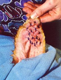
1. The blood is removed from the pinna. This is accomplished by making a small incision in
each end of the hematoma. A drain tube is passed through the hematoma and sutured to the ear.
This assures drainage of any more blood or serum that accumulates in the area. Alternatively, the
skin over the hematoma may be incised and opened completely. This is more likely to be used for
more serious hematomas and for those in which the blood has clotted.
2. The space where the blood accumulated is obliterated. Since the skin over the hematoma
has been pushed away from the cartilage, it must be reattached to it to prevent another hematoma
from occurring. This is accomplished by a series of sutures that are passed through the ear flap.
3. The pinna is stabilized to prevent further damage. The presence of the drain tube will cause
the cat to shake its head even more. Shaking at this time may cause further damage to the pinna.
Therefore, the pinna is laid on top of the cat's head and bandaged in place. Although the bandage
may be somewhat cumbersome, it will prevent further damage to the pinna and allow proper healing
to progress.
4. The cause of the problem is diagnosed and treated. Another important aspect of treatment
is dealing with the cause of the shaking. If an infection is present, medication is prescribed to treat
the problem. However, some cats have no infection but have foreign material lodged in the ear
canal such as a tick, piece of grass, etc. Any foreign material is removed. It is also possible that a
foreign body initiated the shaking but was later dislodged. If that occurs, and no infection is present,
further treatment of the ear canal is not needed.
What follow-up treatment is needed?
The drain tube and bandage are generally removed in about 3-14 days. At that time, the hematoma
is usually healed. There will be two holes in the skin where the drain tube entered. They will close
within a few days. If discharge occurs from the holes before they close, it should be cleaned off with
hydrogen peroxide. In some cats, the stitches through the ear flap will be removed, and in others
they dissolve.
If an infection was present, it will be necessary to recheck the ear canal to be sure that the infection
is gone. Otherwise, another hematoma may occur.
An aural (ear) hematoma is a collection of blood, serum, or a blood clot within the pinna (ear flap).
When present, the pinna will appear very thick and spongy. The swelling may involve the entire
pinna or it may involve only one area.
How is it caused?
When something irritates the ear canal, the cat responds by scratching or shaking its head.
Excessive shaking causes blood vessels to break, resulting in bleeding. An understanding of the
ear's anatomy makes the sequence of events more logical.
The ear flap is composed of a layer of skin on each side of a layer of cartilage. The cartilage gives
the ear flap its shape. Blood vessels go from side-to-side by passing through the cartilage. Violent
shaking causes the vessels to break as the skin slides across the cartilage.
What is the treatment?
There are two approaches used to treat aural hematomas. The first is the conservative approach. A
needle is used to withdraw the fluid from within the pinna, and an injection of a corticosteroid is
made into the area that contained the fluid. The pinna is bandaged so that pressure is applied to it
to prevent recollection of fluid. This method is used when the hematoma is small or if financial
limitations prevent surgery. However, the success rate is less than 50% and frequently result in
severe scarring.
Because the success rate is so low with conservative treatment, most ear hematomas are treated
with surgery. If surgery is chosen, there are four commonly used steps. However, different situations
require different surgical techniques.
1. The blood is removed from the pinna. This is accomplished by making a small incision in
each end of the hematoma. A drain tube is passed through the hematoma and sutured to the ear.
This assures drainage of any more blood or serum that accumulates in the area. Alternatively, the
skin over the hematoma may be incised and opened completely. This is more likely to be used for
more serious hematomas and for those in which the blood has clotted.
2. The space where the blood accumulated is obliterated. Since the skin over the hematoma
has been pushed away from the cartilage, it must be reattached to it to prevent another hematoma
from occurring. This is accomplished by a series of sutures that are passed through the ear flap.
3. The pinna is stabilized to prevent further damage. The presence of the drain tube will cause
the cat to shake its head even more. Shaking at this time may cause further damage to the pinna.
Therefore, the pinna is laid on top of the cat's head and bandaged in place. Although the bandage
may be somewhat cumbersome, it will prevent further damage to the pinna and allow proper healing
to progress.
4. The cause of the problem is diagnosed and treated. Another important aspect of treatment
is dealing with the cause of the shaking. If an infection is present, medication is prescribed to treat
the problem. However, some cats have no infection but have foreign material lodged in the ear
canal such as a tick, piece of grass, etc. Any foreign material is removed. It is also possible that a
foreign body initiated the shaking but was later dislodged. If that occurs, and no infection is present,
further treatment of the ear canal is not needed.
What follow-up treatment is needed?
The drain tube and bandage are generally removed in about 3-14 days. At that time, the hematoma
is usually healed. There will be two holes in the skin where the drain tube entered. They will close
within a few days. If discharge occurs from the holes before they close, it should be cleaned off with
hydrogen peroxide. In some cats, the stitches through the ear flap will be removed, and in others
they dissolve.
If an infection was present, it will be necessary to recheck the ear canal to be sure that the infection
is gone. Otherwise, another hematoma may occur.

Animal Hospital of Fate 1001 North W.E.Crawford (Highway 66 in Fate) Rockwall, Texas 75087 972-722-0066 |
| Rockwall, Texas 75087 972-722-0066 |